-
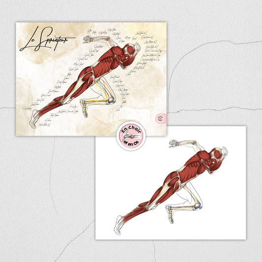
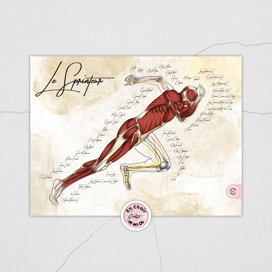
Anatomical chart of the sprinter - Usain Bolt
Regular price From €25,00 EURRegular priceUnit price per -
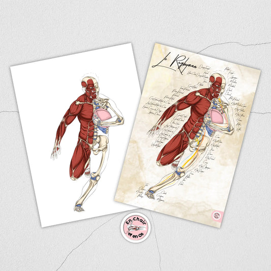
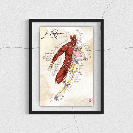
Rugby anatomical chart
Regular price From €25,00 EURRegular priceUnit price per -
Basic anatomical charts pack
Regular price From €35,00 EURRegular priceUnit price per€50,00 EURSale price From €35,00 EURSale -
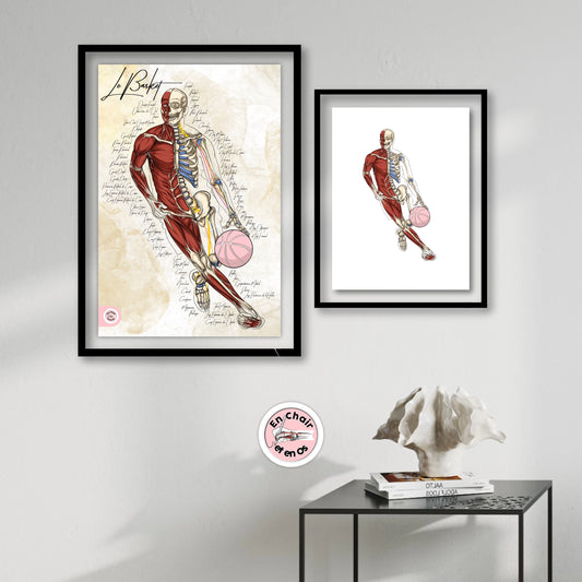
Tony Parker Basketball Anatomical Poster
Regular price From €25,00 EURRegular priceUnit price per -
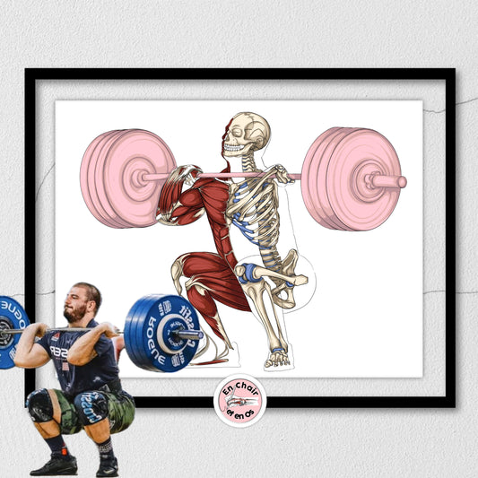
Anatomical weightlifting poster - The squat
Regular price From €25,00 EURRegular priceUnit price per -
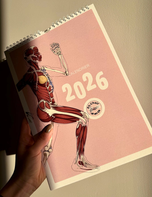
2026 Calendar – Anatomy in Motion
Regular price €18,00 EURRegular priceUnit price per€35,00 EURSale price €18,00 EURSale -
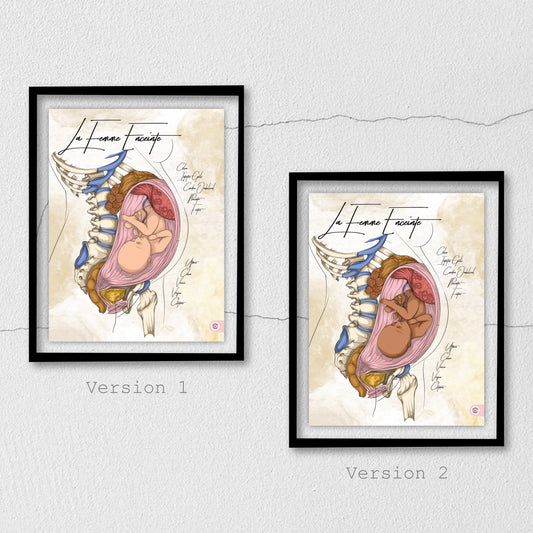
Anatomical chart of a pregnant woman
Regular price From €59,00 EURRegular priceUnit price per -
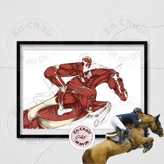
Anatomy of the horse - Anatomical chart for the rider
Regular price From €25,00 EURRegular priceUnit price per -
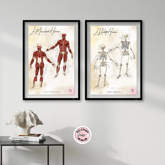
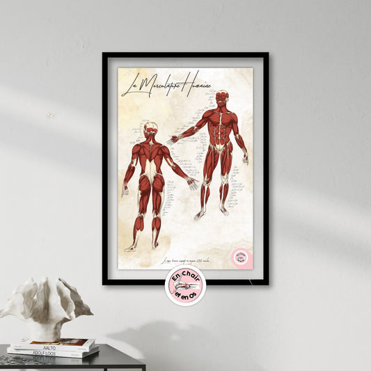
Anatomical chart of human musculature
Regular price From €27,00 EURRegular priceUnit price per -
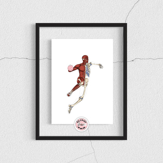
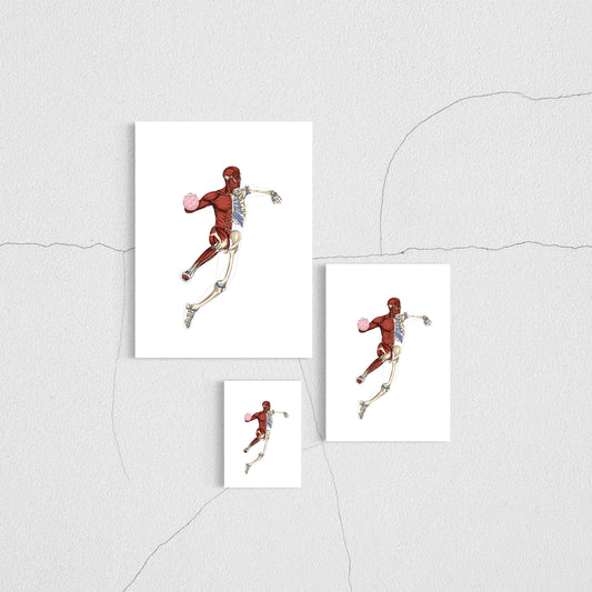
Anatomical flayed model of a handball player
Regular price From €25,00 EURRegular priceUnit price per -
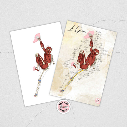
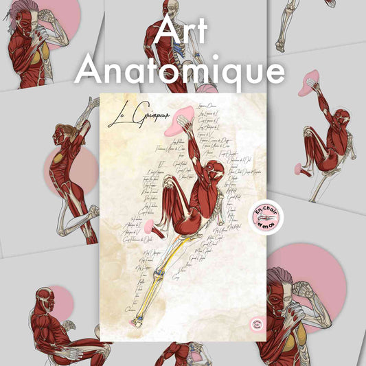
Anatomical chart of rock climbing
Regular price From €25,00 EURRegular priceUnit price per -
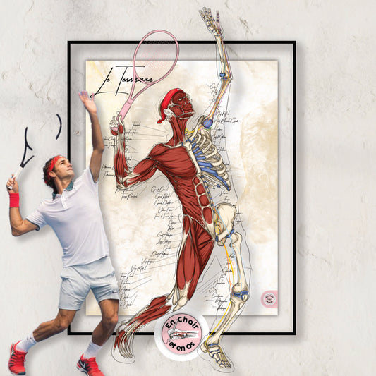
Anatomical chart of the tennis player - Roger Federer
Regular price From €25,00 EURRegular priceUnit price per -
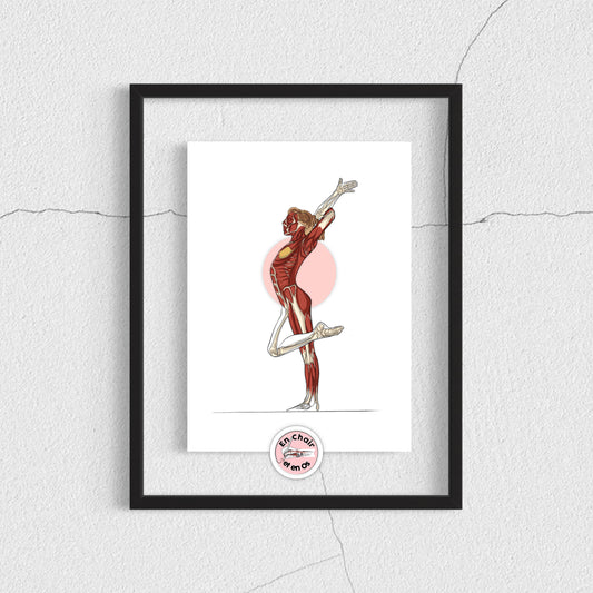
Anatomical flayed figure of a gymnast
Regular price From €25,00 EURRegular priceUnit price per -
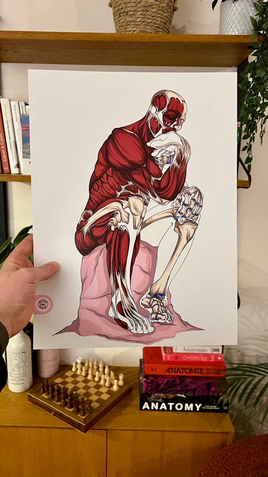
Anatomical flayed figure of Rodin's The Thinker
Regular price From €25,00 EURRegular priceUnit price per -
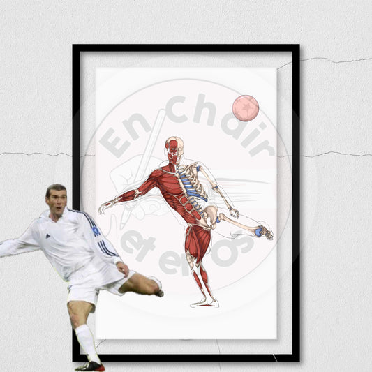
Anatomical Football Poster - Zidane
Regular price From €25,00 EURRegular priceUnit price per -
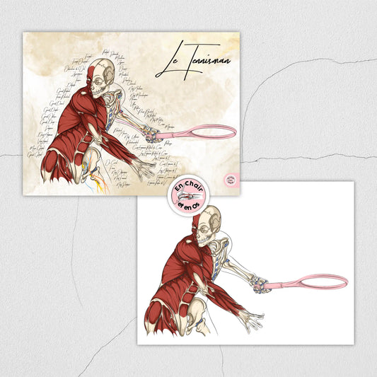
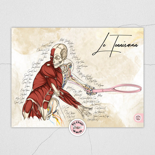
Anatomical chart of a tennis player
Regular price From €25,00 EURRegular priceUnit price per -
Anatomical dance poster
Regular price From €25,00 EURRegular priceUnit price per€22,00 EURSale price From €25,00 EUR -
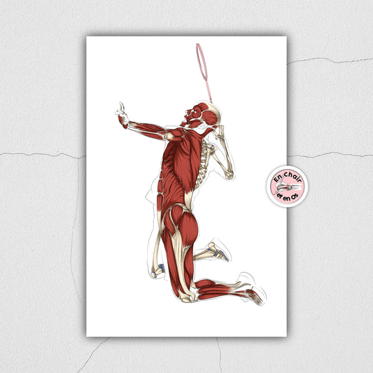
Anatomical flayed model of badminton
Regular price From €25,00 EURRegular priceUnit price per -
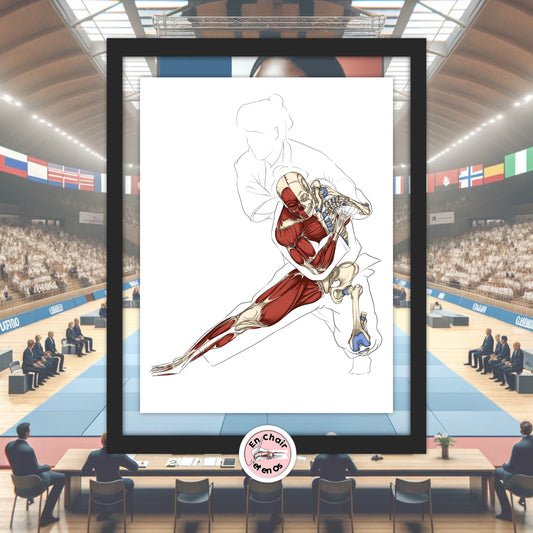
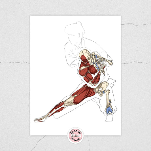
Anatomical Flayed Judo Figure
Regular price From €25,00 EURRegular priceUnit price per -
Anatomical chart of the human skeleton
Regular price From €27,00 EURRegular priceUnit price per -
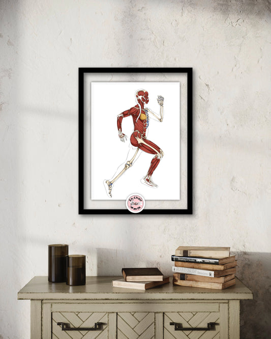
Anatomical flayed figure of running - the marathon runner
Regular price From €25,00 EURRegular priceUnit price per -
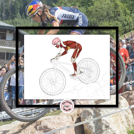
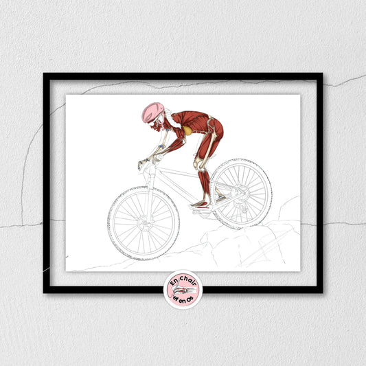
Anatomical cutaway of a mountain bike
Regular price From €22,00 EURRegular priceUnit price per -
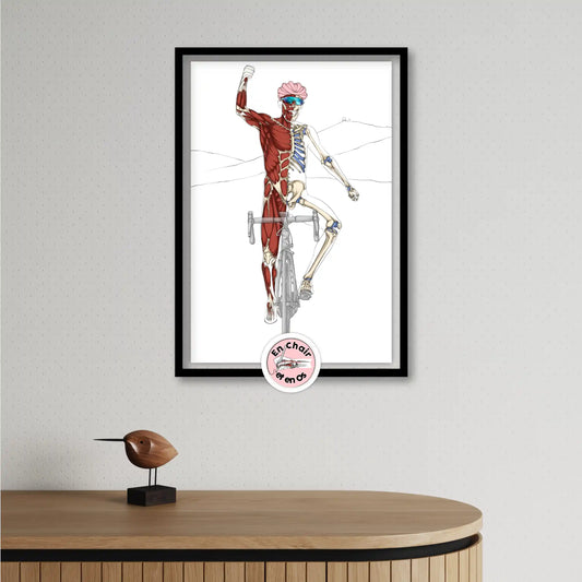
Thibaut Pinot Cycling Anatomical Flayed Figure - Tourmalet Victory
Regular price From €25,00 EURRegular priceUnit price per€0,00 EURSale price From €25,00 EUR -
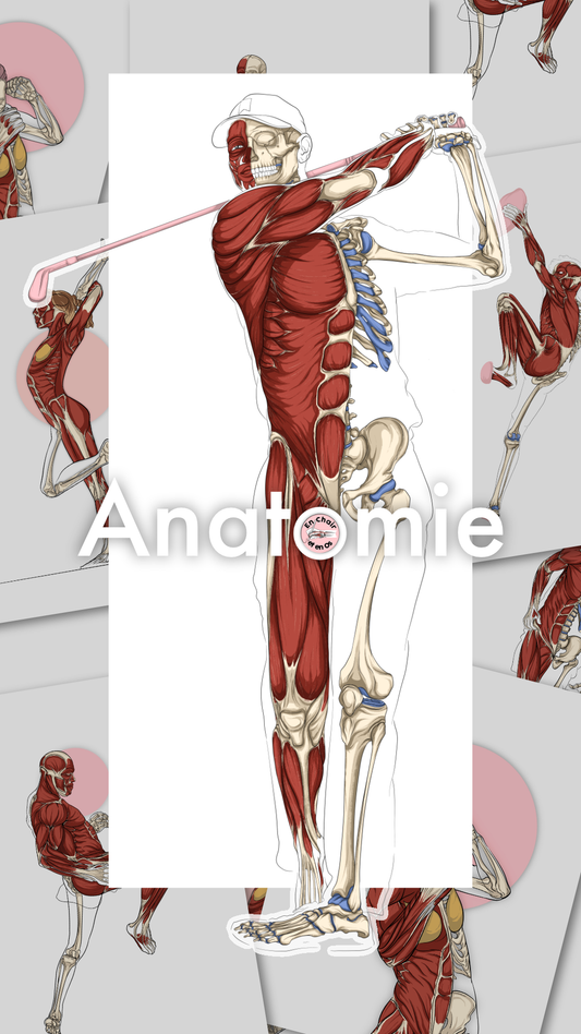
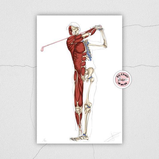
Golfer's Anatomical Flayed Figure - LIMITED EDITION
Regular price From €50,00 EURRegular priceUnit price per














Collection: Flesh and Bone anatomical plates
Anatomical charts of the human skeleton
Anatomical poster of the spine
Human anatomy is often illustrated by the skeleton, as seen on the anatomical skeleton chart, and thus by the spine, shown on the various anatomical skeleton charts. The spine, or "rachis," is composed of:
- 7 cervical vertebrae,
- 12 thoracic vertebrae,
- 5 lumbar vertebrae,
- fused sacral vertebrae that form the sacrum and end with
- the coccyx.
Which you will find on the various En Chair et en Os anatomical charts.
At the thorax level, the spine is accompanied by the costal cage, 12 pairs of ribs, attached at the front to the sternum by sterno-costal cartilages; they are all visible on the psoas-iliac anatomical chart.

Anatomical poster of the skull
The skull is a group of body bones that are regularly seen in medical offices, osteopathy offices, or physiotherapy clinics. It is composed of paired and unpaired bones: the frontal, parietal, temporal, occipital, maxillary, mandible... which can be found on the anatomical chart of skull bones.
Inside the skull are the structures present on the anatomical chart of the reciprocal tension membranes, with the falx cerebri, the tentorium cerebelli, the pituitary gland, and the falx cerebelli, as depicted on the anatomical chart of the cranial diaphragm, which is part of an osteopathic concept.

Anatomical chart of the arms
Depending on the setting, the need to show a model of the anatomy of the shoulder skeleton, arm, elbow, or hand may be necessary, such as for students in medicine, manual therapy, but also art.
The upper limb includes the shoulder area, with the scapula, clavicle, and humerus. The shoulder has 5 joints: 3 "true" and 2 "false" ones.
The elbow articulates further down with the radius and the ulna to form the elbow. The ulna and radius articulate further down with the carpal bones to form the model of the hand with the metacarpals and phalanges.
The anatomical chart of arm muscles summarizes all the muscles of the arm.

Anatomical poster of lower limbs
The study of human and medical sciences also shows us the importance of learning human anatomy, which includes the lower limb. This includes the hip, formed by the coxal bone and the femur, providing the coxofemoral joint. The femur articulates further down with the tibia, fibula, and patella to form the knee, the femorotibial and superior tibiofibular joints.
Further down, the tibia and fibula articulate with the talus to form the ankle. The foot is composed of tarsal bones, ankle bones, and phalanges.
The anatomy of the leg is shown on the various anatomical charts of leg muscles and the anatomical charts of the leg skeleton.
Anatomical chart of the human body muscles
Anatomical poster of leg muscles
The leg model includes various groups of muscles, as depicted on the anatomical chart of leg muscles from En Chair et en Os, connecting the hip to the knee. The main groups are:
-
The quadriceps, located at the front of the leg surrounded by the sartorius, with:
-
The rectus femoris
-
The vastus lateralis
-
The vastus medialis
-
The vastus intermedius
-
The hamstrings at the back of the leg, with:
-
The biceps femoris
-
The semimembranosus
-
The semitendinosus
-
The adductors, inside the thigh, with:
-
The adductor brevis
-
The adductor longus
-
The adductor magnus
The gracilis and pectineus accompany the adductors.
All these muscles are uniarticular or polyarticular and allow for flexion, extension, adduction, or abduction movements of the leg under the thigh and hip.
Other muscle groups span from the leg to the foot and are represented on the various anatomical charts from En Chair et en Os.

Anatomical chart of arm muscles
The arm includes various muscle groups, all represented on the anatomical chart of arm muscles.
On a human model, what stands out most often on an arm is the biceps and triceps brachii. However, all muscles play their role in the proper physiology of the arm. Hence the importance of the functionality of the rotator cuff, composed of:
-
Teres minor
-
Supraspinatus
-
Infraspinatus
-
Subscapularis
-
(long head of biceps brachii)

Anatomical poster of abdominal muscles
-
The abdominal wall includes 4 major muscle groups:
-
The rectus abdominis
-
The external obliques
-
The internal obliques
-
The transversus abdominis
All are represented on the anatomical chart of trunk muscle chains made by En Chair et en Os.

Anatomical chart of foot muscles
The foot is represented on the foot charts, hand-drawn by En Chair et en Os.

Anatomical chart of the perineum
The anatomical representation of the perineum shows its importance in both women and men, particularly regarding variations in intra-abdominal pressure, as well as quality of life (sexuality, bowel movements, urination...).
It can be synthesized as the pelvic diaphragm, though this does not include all the muscles of the perineum.

Anatomical chart of the nervous and vascular system
Anatomical poster of the brain
The brain is often represented in human sciences; it provides beautiful decoration for various medical, psychotherapy, psychiatry, chiropractic, and osteopathy offices.
Anatomical chart of nerves
Nerves are the source of movement and the transmission of nervous information. They can be represented by the central nervous system (brain + spinal cord) but also by the autonomic nervous system.
Anatomical representation via a chart of the nervous system is recurrent in order to provide a clear visual of their endings and actions, such as on the anatomical chart of Rodin's The Thinker.
The autonomic nervous system can also be depicted with its various ganglia and relationships to viscera and organs.

Anatomical chart of organs
Anatomical poster of the heart
The heart is a key element, especially when discussing pathology. The heart is the source of the proper functioning of the entire organism, as it circulates blood permanently and regularly throughout the body. It can be a beautiful element of anatomical decoration in an office.
Anatomical chart of the lungs
The lungs are key to the oxygenation of the human body and part of the respiratory system; accompanied by other elements, they can be a beautiful decoration of the respiratory system.
Anatomical poster of the digestive system
The digestive tract, by En Chair et en Os, is the key to digestion and thus the absorption of elements essential for the proper functioning of the human body. It draws its energy from food: lipids, carbohydrates, and proteins.

Anatomical chart of the genitourinary system
The kidneys, ureters, bladder, and urethra allow for showing the entire system that can be affected by pathology.
