Human Anatomy: Understanding the Body with Images and Diagrams – In Flesh and Bone
What if your body were not a mystery, but a readable system, a structure made of units that cooperate? Under the skin, everything is composed: a cell becomes tissue, tissue becomes an organ, the organ takes on a role and a function. Blood flows through the body in its vessels, muscles contract for movement, nerves and the nervous system send signals to the hand, the lower limb, the head, and the neck. And suddenly, health takes on concrete meaning: breathing, circulatory system, muscular system, digestive system... everything becomes a map.

What fascinates me is that everything is connected. The body is not a list of spare parts. It is composed of units (cells, tissues, organs) that communicate and adapt. Systems organize themselves to ensure essential functions: breathing, nourishing, moving, repairing, adapting. And behind this mechanics, there is also a simple logic: understand the form to understand the gesture, understand the structure to understand the role, understand the detail to understand the whole.
Recurrent questions about anatomy are:
- How to learn anatomy and, above all, how to truly understand it (and not just recite it for an exam)?
- What are the different branches and types of anatomy, depending on whether one is taking a medical, sports, artistic, or “general knowledge” approach?
- How to find beautiful anatomical charts, a clear fact sheet, a good anatomy manual, a complete anatomy guide, or a human anatomy book that helps with visualization?
To give a definition of anatomy, it is the study of the structure and systems that make up the human body. It is a foundation for learning and understanding in medical fields, sports, but also art, with morphological or artistic anatomy. And what I love is that anatomy is never “disconnected from reality”: as soon as one studies it seriously, one touches upon human physiology, that is to say, living, dynamic, adaptive functioning. We move from “where it is” to “what it’s for” and “how it works”.
In this article, I will explore the major regions of the body – from head to trunk, from upper limb to lower limb – and dive into major systems: muscular, nervous, vascular, cerebral, pulmonary, renal, digestive, cardiac. I will also treat the body by zones: head, neck, thorax, abdomen, pelvis, because that is often what brings the most clarity to explanations: “in this zone, what contains what, what passes through, what moves?”.
Finally, I will address two key questions: how to learn and understand anatomy? And how to transform this knowledge into something concrete in your daily life as a practitioner, student, or artist: images, diagrams, a video, 3D resources, methods, and also artworks to display, because yes: learning anatomy, understanding it, and sometimes hanging it on the wall is also a way of living it.
With anatomical illustrations, written content, definitions, free courses, and educational aids, En Chair et en Os supports you during your studies to better visualize and learn, and then in your professional life to give an identity to your spaces: practice, gym, studio, office. I often say it: a well-thought-out chart or fresco can be both aesthetic, educational, and memorable.
What is human anatomy?
Definition of anatomy
The definition of anatomy: anatomy is the science that studies the structure of organized living beings. Human anatomy, in particular, describes the form, arrangement, and relationships of the organs and tissues that constitute the body. In other words, it answers questions like:
- Where is a structure located?
- What is it related to?
- Where do the nerves and vessels pass?
- What protects what, what supports what?
Anatomy is indispensable for understanding the body in its normal state (physiological), but also its alterations: trauma, inflammation, wear, degeneration, or disease. This link between “normal” anatomy and clinical situations is what makes the discipline essential in medical fields. It is also what makes anatomy fascinating: we move from diagram to life.
Historically, anatomy relied on the dissection of cadavers, a foundational practice. Today, it also benefits from imaging tools (X-rays, CT scans, MRI, ultrasound) which allow for the exploration of the living in a precise, non-invasive way adapted to the study of the organism in motion. These tools do not replace dissection, but they change the way of learning: we see anatomy “over time,” in its volumes, and sometimes at different levels (slice, 3D reconstruction, etc.).
Anatomy, biology, and the cell: the common thread
We sometimes forget that between the large anatomical chart and reality, there is a continuity: the structure of the body comes from cellular organization. A muscle cell does not have the same shape or the same function as a nerve cell. A cell of an epithelial tissue (epidermis) does not look like a cell of connective tissue. And yet, they all integrate into organs, then into systems. Understanding this is already building a bridge between anatomy, biology, and physiology.
Origin and etymology of the word “anatomy”
The word “anatomy” comes from Ancient Greek anatomein (ana- = through, temnein = to cut), literally “cutting through.” This etymology reminds us that the discipline was born from dissection, used since Antiquity to reveal the internal structure of the body. It is raw, but it is clear: we cut, we observe, we describe, we compare, we draw. And above all: we learn.
The great names in the history of human anatomy
When talking about human anatomy, certain names often come up (non-exhaustive list):
- Hippocrates (5th century BC): considered the father of medicine, his writings already include anatomical observations and a rational approach.
- Galen (2nd century): Roman physician, he established major anatomy treatises based on animals, influencing medicine for over 1,000 years.
- Andreas Vesalius (16th century): revolutionized anatomy with his De Humani Corporis Fabrica (1543), thanks to study through rigorous dissection.
- Leonardo da Vinci (15th-16th century): his anatomical drawings, blending art and science, remain among the most detailed and inspiring.
What I love about this history is that it shows something important: anatomy advances when we agree to look, verify, correct, and represent clearly. That’s also why I make images: because a good image can shorten a week of misunderstanding.
But who is Andreas Vesalius?
Andreas Vesalius (1514-1564) is considered the father of modern anatomy. A Brussels-born physician and professor, he transformed the knowledge of the human body in the 16th century by performing dissections on cadavers himself, which was exceptional at the time.
His major work, De Humani Corporis Fabrica (1543), illustrated with anatomical charts, marks a break with the ancient theories inherited from Galen. Vesalius demonstrated that many descriptions of animal anatomy had been wrongly applied to humans. His work founded a visual approach to anatomy, and this approach remains a reference today.
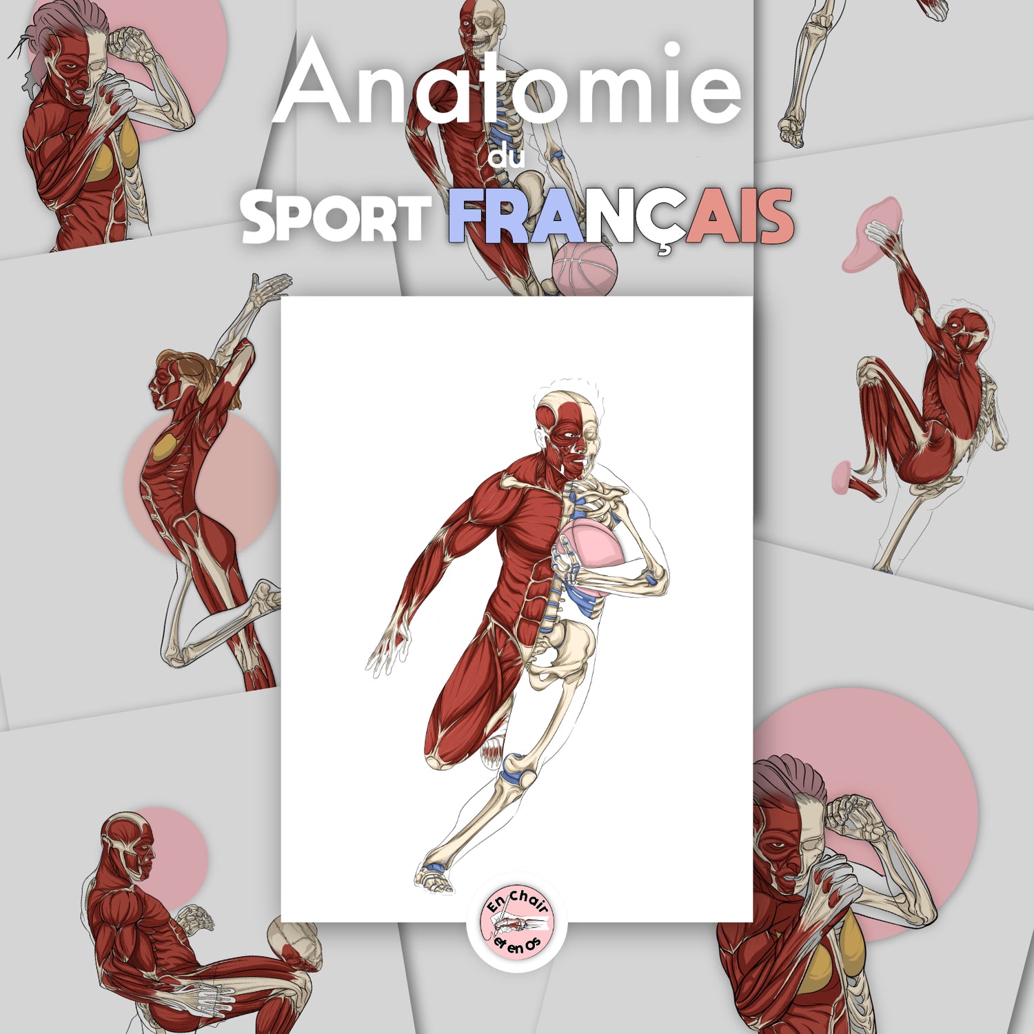
It was upon discovering these works that I had the idea to make a collection on anatomical flayed figures in sports.
The great authors of descriptive anatomy treatises still studied today
Besides Vesalius, several authors have left a lasting mark on the history of descriptive anatomy, and their works remain references in education. In physiotherapy studies in France, some manuals are widely used, and schools often have their preferences.
- Henry Gray (1827-1861): his famous Gray’s Anatomy, published in 1858, is still consulted worldwide.
- Jean Cruveilhier (1791-1874): French professor, author of a monumental treatise on descriptive anatomy accompanied by detailed plates.
- Léo Testut (1849-1925): French physician whose human anatomy treatise remains a classic.
- Frank Netter (1906-1991): American illustrator and physician, his illustrated anatomical atlases have become essential.
- Rouvière, Brizon, Castaing, Baqué… and others.
These authors share the fact that they produced very visual and educational works. For a student, a good book is not the one that “contains everything.” It is the one that provides a common thread, clear vocabulary, and a structure that allows for effective review.
The different studies and branches of anatomy
Depending on your teacher and your curriculum, you will go more or less deep. Some courses focus on the basics (bones, muscles, joints). Others address all systems, with varying levels of detail.
Anatomy can be approached in two ways:
- by system (respiratory, nervous, digestive…)
- by zone (shoulder, hand, head and neck…)
Both approaches are useful. The first provides a functional logic. The second provides a spatial logic. And in practice, to learn quickly and well, one often ends up mixing both.
Anatomy of the human body's systems
What is a system?
A system is a set of organs and tissues that work together to fulfill a precise function. For example, the respiratory system ensures gas exchange: it allows for the supply of oxygen and the elimination of carbon dioxide.
Anatomical study by system consists of describing each one separately, highlighting their organization, their relationships, and their connections with other systems.
The main systems (clear list)
Here is the list of systems. Over time, I will offer illustrations, animations, and written support, page by page, to make all of this accessible:
- Skeletal system: bone structure that supports the body and protects organs. The axial skeleton is a major landmark for orientation.
- Muscular system: muscles allowing for movement and posture. Muscle tissue contracts, pulls on bones, stabilizes joints.
- Nervous system: central (brain, cord, brainstem) and peripheral. It receives, processes, and returns signals.
- Cardiovascular system / circulatory system: heart and vessels. It transports blood, nutrients, and participates in regulation.
- Respiratory system: lungs, trachea, bronchi. It manages O₂ / CO₂ exchanges.
- Digestive system: from the oral cavity to the digestive tract (stomach, intestines…) with liver, pancreas. It transforms and absorbs.
- Urinary system: kidneys, ureters, bladder. It filters, eliminates, and regulates water and certain internal parameters.
- Genital system / reproductive system: reproduction, hormones, continuation of the species. We will detail the female reproductive system (ovaries, tubes, uterus).
- Endocrine system: hormonal glands, regulation of major balances.
- Lymphatic system and immune system: lymph nodes, lymphatic vessels, defense.
- Integumentary system: skin, nails, hair, protection.
- Sensory system: vision (eye), hearing, smell, taste, touch.

I know this list may seem long. But it becomes simple when you relate it to concrete situations: breathing during exercise, digestion and energy, nerve pain, circulation, recovery, adaptation. That is where anatomy becomes alive.
Human anatomy by zone: learning by regions
Another way to study anatomy is the approach by zones or regions. Systems are grouped according to their location, which allows for analyzing all the structures present in the same region of the body.
For example, the head and neck region includes:
- the bones of the skull,
- the muscles of the face,
- cranial nerves,
- arterial and venous vessels,
- sensory organs (including the eye, nose, mouth, etc...),
- and essential deep structures.
Next, we move down:
- thorax: rib cage, lungs, heart, large vessels
- abdomen: digestive tract, liver, kidneys, etc.
- pelvis: pelvic organs, attachment zones, movement hinge

This regional reading has one advantage: we understand what passes through, what overlaps, and what creates symptoms according to the zones.
At En Chair et en Os, I favor a hybrid approach:
- Studied by zone: muscular, skeletal, peripheral nervous, and arteriovenous systems.
- Studied by system: endocrine, digestive, respiratory, immune, etc.
The 4 types of anatomy (the ones I will mainly use here)
Descriptive anatomy
Descriptive anatomy consists of describing each organ, tissue, or structure separately, without necessarily focusing on their function. It is the oldest approach and the foundation of theoretical learning. It allows for the establishment of a common vocabulary and a reliable mental map.
But as I often say: one cannot totally separate anatomy and physiology. Because a structure exists to perform a function. And a function modifies the structure (adaptation, growth, aging).
Functional anatomy
Functional anatomy links structures to their functions: movement, mechanical constraints, daily gestures, and further, the modifications necessary to perform in sports, or to support patients in their rehabilitation.
It is particularly used in sports medicine, physiotherapy, osteopathy, and manual therapies. This is also where images are powerful: an arrow on a diagram can immediately make one understand an axis, a traction, or a stabilizing role.
Pathological and clinical anatomy
This branch studies the modifications of organs and tissues in diseases (infections, tumors, degeneration...). It allows for the link between normal anatomy and clinical signs.
In this blog, I will mainly detail pathological and traumatic anatomy: fractures, ligamentous injuries, vascular injuries. Understanding a pathology does not mean diagnosing oneself. It means gaining knowledge and explanatory capacity.
Artistic anatomy
Artistic anatomy focuses on the visual representation of the human body. It is used by painters, sculptors, and illustrators to render proportions, reliefs, and volumes. Leonardo da Vinci is a major precursor.
En Chair et en Os is strongly based on morphological and artistic anatomy: it allows me to create anatomical models (écorchés) and offer them to you as wall decorations, while remaining consistent with reality.
How to learn anatomy? Simple tools and methods (but not easy)
The importance of anatomy
Anatomy is essential because it conditions your understanding of the body. It is useful for everything: reading movement, explaining pain, understanding recovery, or general knowledge.
The problem: density. There is a lot to memorize and even more to understand. It is long work. And you must choose your level of precision:
- anatomy for "health": necessary base to be a good practitioner
- anatomy for "leisure": understanding the major structures and the global meaning
In any case, you will need a method.
Basic concepts to know
Before the details, you must learn the anatomical language:
- Conventions: anatomical position, midline, orientation (anterior/posterior).
- Color codes: nerves, arteries, veins, etc.
- Planes and axes: sagittal, frontal, transverse.
- Localization terms: superior/inferior, proximal/distal, medial/lateral.
This foundation turns anatomy into a language. And a language must be practiced.
Effective methods: learning faster
Anatomy is not learned by reading passively. You need a strategy:
- drawing
- simple diagrams
- spaced repetition
- flashcards
- 3D resources
- video
- quizzes
My personal preference is drawing, because it forces you to understand. But everyone must find their own way.
Example of learning: anatomy of the rib cage (thorax abdomen)
To accompany a project like the thorax, I will proceed as follows:
- Descriptive anatomy of the bony elements.
- Addition of muscles, their insertions, their directions.
- Addition of organs: lungs, and especially the heart (cardiac function).
- Addition of nerves and vessels.
- Then: simplified diagram, reproducible drawing, and exercises.
Then, we connect the thorax to the abdomen to understand the continuity: diaphragm, pressures, respiration, posture, links with movement.
Anatomical decorations: the trademark of En Chair et en Os
At En Chair et en Os, I offer several media:
- Posters and anatomical charts: this is the foundation, my trademark.
- Wall murals: a unique practice, a strong identity.
- Anatomical stationery: notebooks, calendars, bookmarks.
In a medical, paramedical, or sports practice, decoration does more than "look nice":
- It creates a reassuring environment.
- It enhances your expertise.
- It becomes a pedagogical tool: explaining with an image is often simpler.

Conclusion: anatomy must be learned, understood, and then displayed on the wall!
Anatomy is a passion for me. It might become one for you too thanks to what I share here. It is forgotten and relearned, again and again. But each time, we understand faster and better.
Thanks to this blog, you will find:
- free flashcards,
- diagrams,
- learning methods,
- visual aids,
- and solutions to decorate your office.
See you very soon,
Axel