

-
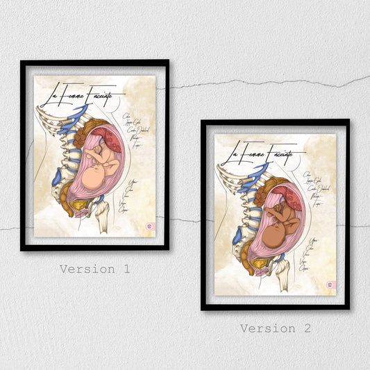
Anatomical chart of a pregnant woman
Regular price From €59,00 EURRegular priceUnit price per

Collection: Anatomical plates of the pregnant woman
The pregnant woman's uterus
What is the anatomy of the pregnant woman's uterus?
As shown on the anatomical chart of the pregnant woman, the uterus is posterior to the vagina. Before pregnancy, the uterus is a hollow, inactive smooth muscle. Unlike the ovaries, the uterus is located below the peritoneum, regardless of the stage or month of pregnancy. It is situated in the pelvic cavity, between the two iliac bones. The female pelvis is wider than the male pelvis, which allows for better management of childbirth.

What are the changes in a pregnant woman?
The non-gravid uterus, which is not carrying a baby, is tilted forward. During the future mother's pregnancy, the small pelvis—containing the fetus (the future baby), the placenta, the umbilical cord, and the amniotic fluid—will grow and undergo changes to its body and cervix throughout the months of pregnancy.
It will grow from a few centimeters to an average of 32 cm above the mother's pubis. It will continue to grow throughout the trimesters of pregnancy, taking up more and more space within the abdomen.

The anatomical illustration of the pregnant woman.
The anatomical illustration of the pregnant woman was created to decorate medical and paramedical offices, such as those of gynecologists, midwives, physiotherapists, osteopaths, and all health and movement professionals who support pregnant women during their months of pregnancy to better welcome the future baby. This anatomical chart of the pregnant woman will be perfect for showing what their bodies look like from the inside, thereby helping to better understand and explain the changes in their physiology.
The goal of the anatomical illustration of the pregnant woman is to show her anatomy in detail in order to provide a fun, engaging, and informative visual for patients, but also for health students, including nursing, midwifery, osteopathy, physiotherapy, and any other health-related professions.

The organs
The pelvic organs undergo changes.
The woman's uterus is surrounded by organs. In front is the bladder. Behind is the rectum. During pregnancy, changes in the body and cervix will cause stress on the body, which we will return to. These changes can be the source of discomforts known during pregnancy, which may explain the increase in nocturnal urination. This can cause excess pressure on the bladder, as well as on the attachments of neighboring organs. Thus, traction on the attachments of the bladder and uterus can affect the kinetics of the lumbopelvic spine, which may explain the back pain commonly experienced during pregnancy.
The intestines during pregnancy before the baby's birth
During the first trimester, since the gravid uterus, fetus, and placenta are not yet highly developed, the impact on other organs is not yet significant. During the second trimester, the uterus, above the vagina, will develop and push the intestines backward. During the 3rd trimester, as shown in the illustration, the uterus takes up all the space before birth. The pressure exerted on the intestines by the baby can slightly disrupt their proper physiological function. Indeed, constipation and gastroesophageal reflux can occur due to excess pressure on the lower esophageal sphincter.
The fetus
The fetus in the uterus during pregnancy
As we see in the illustration of pregnancy, before childbirth and the birth of the baby, the anatomy and physiology of the uterus change. Indeed, the formation and development of the placenta are important; this is checked during medical exams. This marks the end of the ovarian cycle. Fetal growth is enabled by the umbilical cord, which is dedicated to its growth.
The anatomy of the placenta allows it to function properly as a nourishing organ for the baby. Its structure allows it to filter the important elements for the proper nutrition of the fetus throughout the pregnancy. The future child will develop based on the mother's diet during the three trimesters.
Medical illustration for patient and student education
Through drawings, and in particular anatomical charts, we can provide a clearer visual for everyone concerned with this illustration. When a patient can visualize their body, for example on the anatomical chart made by En Chair et en Os, there is visibility of the pelvic organs, vagina, bladder, rectum, sacrum, lumbar vertebrae, but also, and above all, the baby in the future mother's belly.
Pregnancy
The duration of pregnancy
Human pregnancy is an event that lasts on average 9 months, 39 weeks, or 41 weeks of amenorrhea—that is, 266 days after conception or 280 days from the first day of the last period (for a regular 28-day cycle). This is unlike other mammals, where pregnancy can be much longer, which explains the lack of maturity of the baby at birth.
Pregnancy time can be counted in weeks of amenorrhea, which corresponds to the absence of periods. The week of amenorrhea starts from the first day of the last period. This amenorrhea is gestational because it is due to pregnancy. In contrast, the week of pregnancy starts from the assumed date of fertilization.
This calculation is more precise because it is based on physiological events. It generally corresponds to 2 weeks more than those of pregnancy.
Health during pregnancy
Various health elements are modified and can be disrupted during a woman's pregnancy.
The cardiovascular system: cardiac output increases to meet the needs of the uterus and placenta. Uteroplacental blood flow accounts for 20% of total blood. The average heart rate increases from 70 to 90 beats per minute.
The blood volume will increase in proportion to the heart's output. The number of white blood cells will also increase, and iron requirements will rise to produce more red blood cells.
The urinary system will be affected; indeed, kidney function varies in tandem with heart function, and the filtration rate will also be increased. The change in posture will put stress on the lumbar spine and thus on the position of the kidneys.