Author: Naël El Kastani
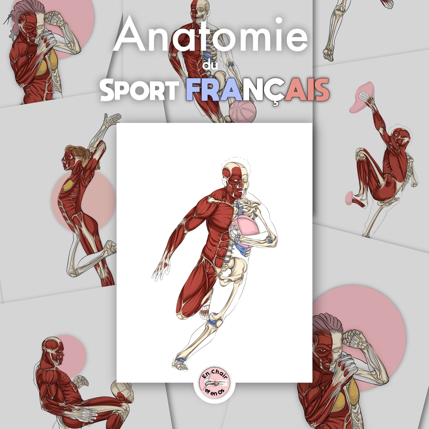
Human anatomy is the foundation of our clinical practice. It is our common language, our shared reference. Yet, in the dynamics of a consultation, using it as a visual tool remains too rare. Far more than a simple illustrative aid, an anatomical diagram is a tool for mediation, precision, and therapeutic support. To concretely illustrate the use of these representations, available on our website En Chair et en Os, they allow for the following in a medical setting:
-
Enriching the caregiver-patient relationship,
-
Strengthening clinical understanding,
-
Facilitating a coherent functional approach to care.
Building a therapeutic bond through anatomy
Visualizing together for better understanding: Muscle chains of the lower limb
In many cases, patients have chronic lower limb pain without understanding the true origin of their symptom. They talk about their left thigh, their glute, or diffuse pain while walking. Their perception of the human body is often fragmented, reduced to what hurts.
This is where the Chart: Muscle chains of the lower limb becomes a powerful therapeutic lever.
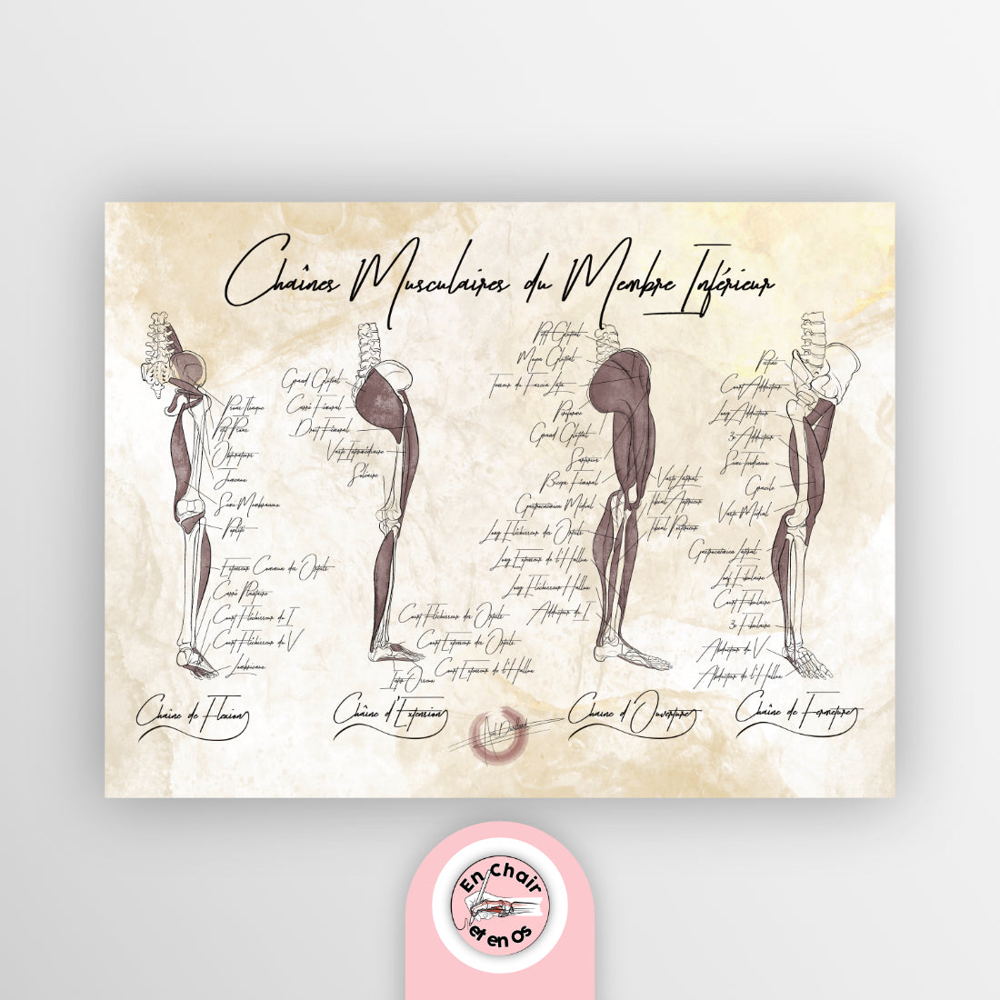
This chart highlights muscle chains, those functional structures that unite several parts of the body. Showing this image to the patient allows them to:
-
Locate the pain within a global muscular system, giving it meaning.
-
Enter into a logic of functional movement, rather than localized treatment.
For the therapist, this information becomes a pedagogical means to explain the link between pain and its biomechanical context. We talk about tissue, network, and function; we start from the surface and move inward.
Providing a clear representation of pain: Pelvic diaphragm
Pelvic pain or functional disorders of the pelvic floor are often complex to address. Without visualization, one hits a wall of misunderstanding or embarrassment.
Using the Chart: Pelvic diaphragm helps break down this barrier.

This diagram highlights the role of the pelvic diaphragm in:
-
supporting organs,
-
venous and lymphatic circulation,
-
respiratory and postural function.
This allows the patient to:
-
grasp that this is not just an intimate area, but a muscular system that acts in coordination with the trunk and the thoracic diaphragm,
-
reclaim their body, with accurate terminology and reference images.
This model becomes a tool for empathetic communication, centered on a functional and living anatomy, rather than a mechanical vision.
Deepening clinical analysis through anatomical images
One brain, one body: Human skeleton
When we talk about general anatomy, we often refer to the human skeleton as a basis. Yet, how many consultations are conducted without ever visualizing this fundamental structure?
The Chart: Human skeleton helps bring order to bodily structure.

In consultation, it:
-
Locates bones, joints, and their relationship with the nervous system and vascular networks.
-
Serves as an anatomical base to explore pain, postural compensations, or movement limitations.
Example: a patient with right shoulder pain and an old pelvic fracture. Showing how the upper limb is suspended from the skeletal trunk via the spine helps anchor understanding in the reality of the human body.
It is also a gateway to physiology and human anatomy, essential for connecting form and function.
Targeting information on muscle function: Human musculature
Which muscle is responsible for which movement? This is a recurring question... but often fuzzy in the patient's mind.
The Chart: Human musculature makes it possible to answer simply by showing each muscle group, their role, their position, and their orientation.

It becomes a tool for clinical precision to:
-
Show the difference between a superficial muscle and a deep muscle.
-
Highlight the action of a rectus muscle, an oblique, or a stabilizing muscle.
-
Explain the impact of a compressed nerve or a loss of muscle strength.
Example: in the clinic, facing a tendinopathy of the upper limb, showing the insertions, muscle synergies, and functional chains helps the patient understand the proposed treatment.
It is also a pedagogical support for trainees or young colleagues: we return to the map of the territory.
Supporting therapeutic strategy through imagery
Linking symptoms and physiology: Human heart
Using the Chart: Human heart allows us to move beyond the vision of an "isolated organ" to evoke an integrated system: vascular system, autonomic nervous system, perfusion mechanisms.

In a consultation where fatigue, palpitations, or peripheral vascular disorders are mentioned, this anatomical diagram allows us to:
-
Locate the organ in the center of the thorax,
-
Show the link between the heart, vessels, and peripheral tissues,
-
Assimilate the relationships with the brain, the hormonal system, and the sympathetic and parasympathetic nervous systems.
This image helps address human functioning as a whole, and link the symptom to its structural logic.
Building a functional project with the patient: The sprinter
The Chart: The sprinter is a dynamic, functional, and integrative representation of the human body in motion.
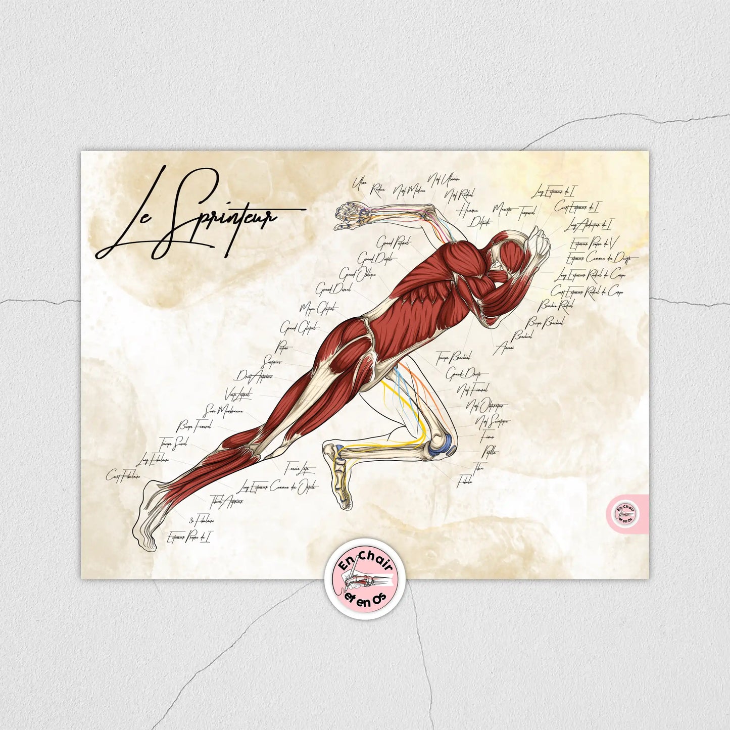
It allows for moving away from a passive logic of pain to enter an active logic of progression:
-
Showing the patient how muscle chains, neuromuscular networks, and joint systems cooperate to produce a functional movement.
-
Giving meaning to rehabilitation, training, or a re-athleticization program.
The organism is seen as a structured and coherent whole, not as an aggregate of independent parts.
This helps establish a long-term vision, oriented toward recovery and autonomy.
Conclusion
These anatomical diagrams help to:
-
strengthen the therapeutic relationship,
-
improve clinical understanding,
-
guide a coherent and dynamic approach to care.
Each chart available on our website En Chair et en Os, offers a valuable, reliable, and pedagogical tool. Using an anatomical diagram in a consultation is not about simplifying. It is about clarifying, making accessible, and giving meaning to complexity.