

Excellente communication avec Axel. D’une gentillesse extraordinaire et efficace. Les visuels sont juste magnifique.
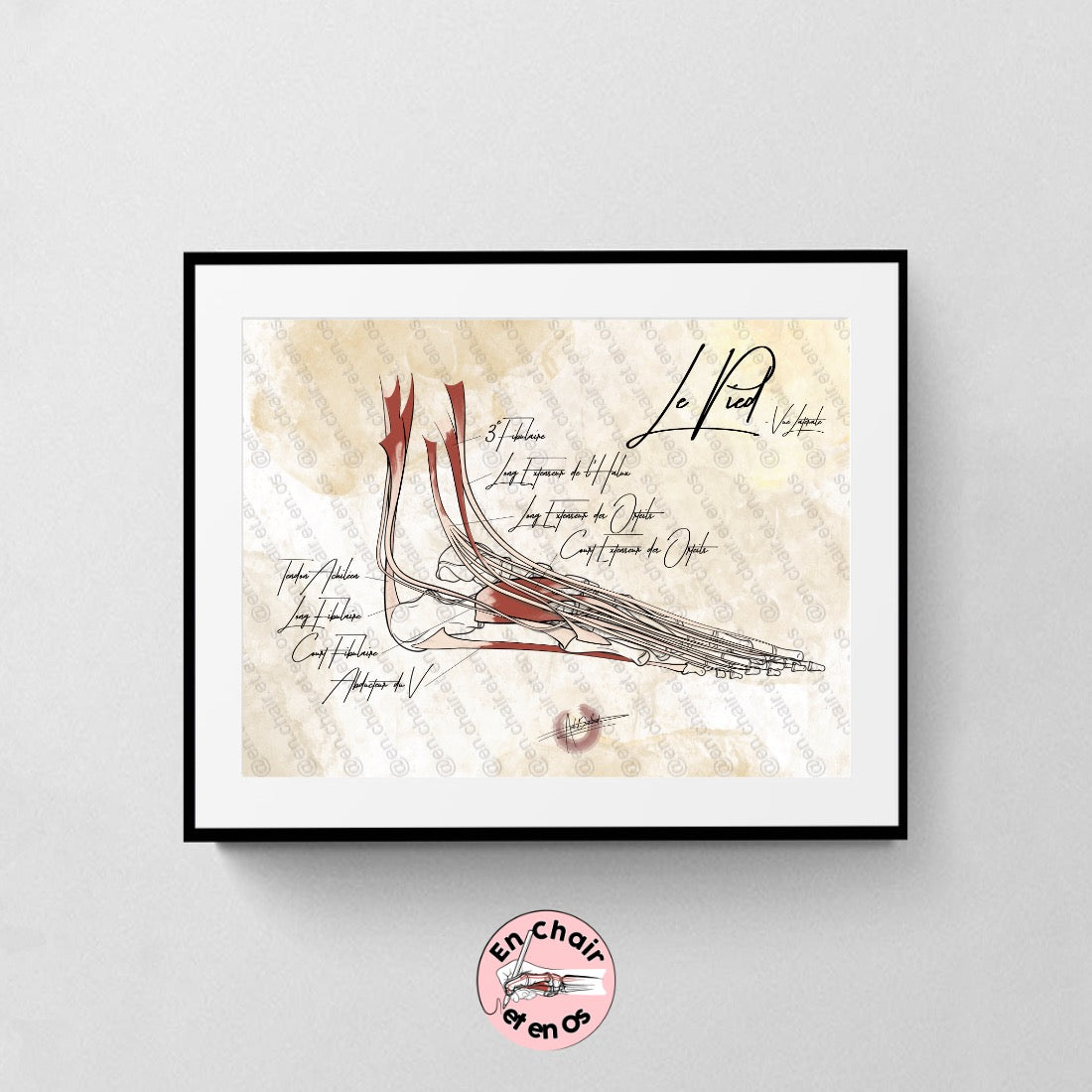
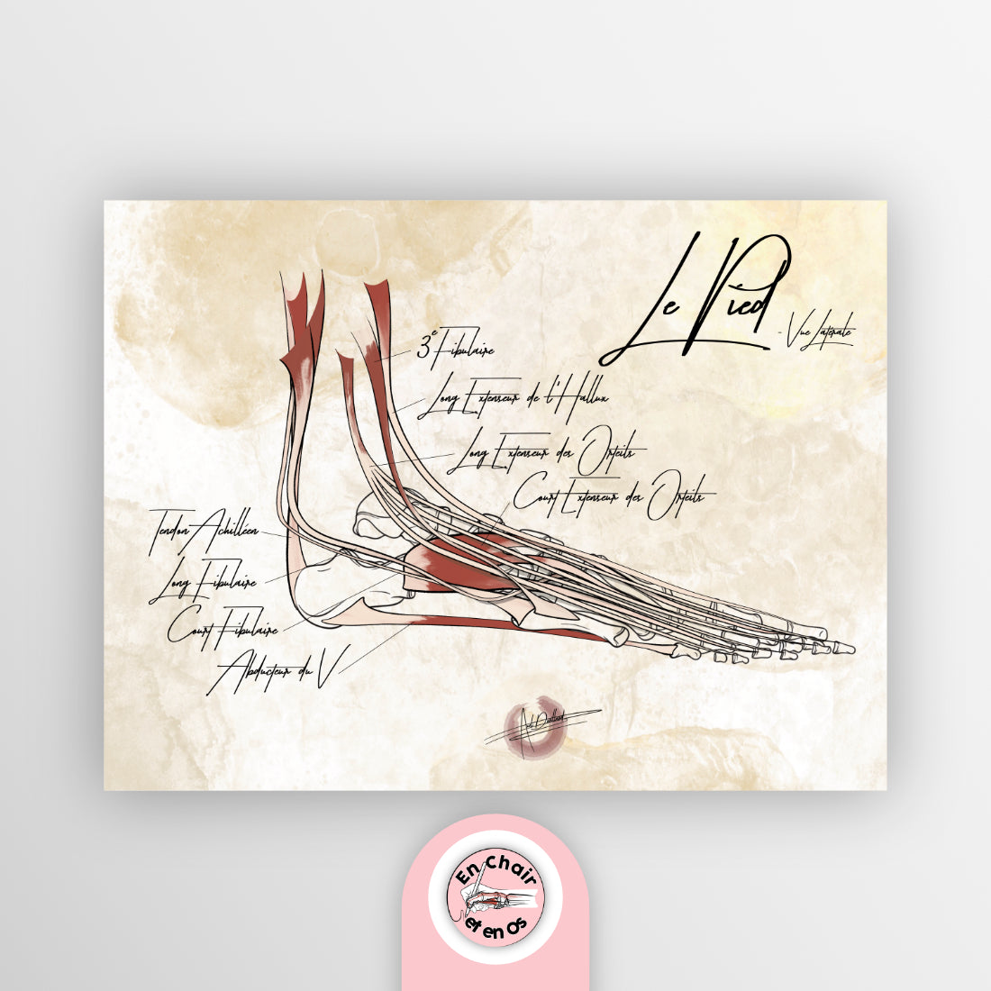
Planche anatomique du Pied - Vue latérale -
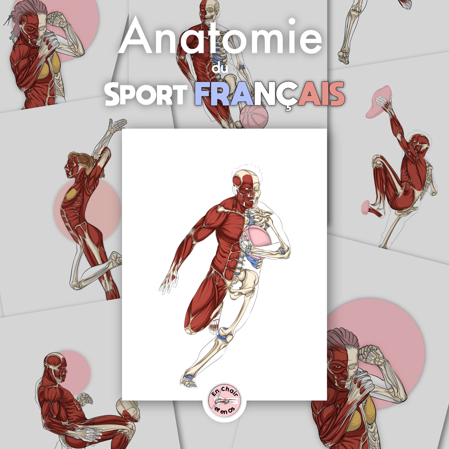
Are you a sports and anatomy enthusiast? You are going to...
En Chair et en Os
Couldn't load pickup availability
Hand-illustrated by Axel Devillard, creator of En Chair et en Os.
The foot bears the entire weight of the body with every step. This anatomical chart of the foot shows it in profile, from the external side, with its bones and muscles hand-drawn. A clear view to understand how the foot articulates with the ankle and how it stabilizes during walking.
An ideal visual aid for explaining an ankle sprain, tendinopathy, or pain on the outer edge of the foot.
The foot has 26 bones: 7 tarsal bones (including the talus and calcaneus), 5 metatarsals, and 14 phalanges. In addition, there are small sesamoid bones under the head of the first metatarsal.
The lateral view shows the profile of the arch, the ankle, and the external edge of the foot, where most sprains occur. It is the most intuitive view for a patient.
The 30 × 40 cm is suitable for a desk or treatment room. The 40 × 60 cm is readable from a distance and becomes the centerpiece of a clinic wall.
Choose your size and receive your anatomical chart of the foot.