Beyond stock images: the demands of a peripheral nerve surgeon
When you type "surgical illustration" into a search engine, you get thousands of results. Vector images or medical stickers under royalty-free licenses. Standardized content, often of good quality, and sometimes even free.
But what does a practitioner do when none of these resources meet their needs? When they are looking for an ultra-precise anatomical illustration to document a complex surgical technique?
This is exactly the situation for Dr. Fernando Davila Fernández, a hand and peripheral nerve surgeon. After months of fruitless searching in image banks, he contacted En Chair et en Os to create something unique.
The challenge: two anatomical illustrations with absolute surgical precision
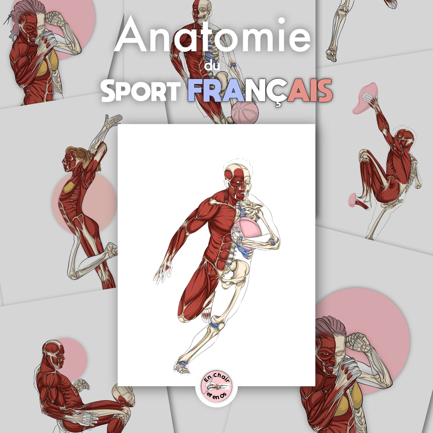
The project was clear: to create two anatomical illustrations intended for medical and educational use. Not a generic drawing found on any stock photo platform. Not a standard medical graphic with simplified icons. But a faithful representation of the ulnar nerve path and an ultrasound-guided surgical technique.
Illustration 1: The ulnar nerve
This first illustration depicts the complete path of the ulnar nerve with surgical precision: Osborne's ligament, the flexor-pronator aponeurosis, the arcade of Struthers, the medial intermuscular septum, and the medial epicondyle. Every anatomical detail counts. Every structure must be positioned exactly as in a real human body.
The unique feature of this illustration lies in the representation of the anatomical continuity between Osborne's ligament and the flexor-pronator aponeurosis. This anatomical detail, often simplified in standard medical illustrations, is faithfully reproduced here based on direct observations from cadavers. The tool thus becomes a valuable surgical reference for understanding zones of nerve compression.

Illustration 2: Ultrasound-guided technique
The second image shows the sectioning of Osborne's ligament under ultrasound guidance. Ultrasound probe, Kemis scalpel, structures visible during the operation: everything is represented. An essential educational tool for training surgery students.
This illustration represents a real technical challenge: combining anatomical clarity with operative elements (probe, surgical instrument) while maintaining optimal readability. The result is an image that can be used both in training and in the operating room as a reference support.

The methodology: when the dissection table becomes a creative studio
Unlike standard vector illustrations created from generic models, this project relies on an exceptional resource: photos of cadaver dissections performed by Dr. Davila himself.
The creation process spanned several months:
- Dissection and photographic documentation: The practitioner performs the dissections and provides high-resolution photos
- Initial draft: First drawing based on the provided images
- Technical feedback loops: Fine anatomical adjustments based on the practitioner's notes
- Final validation: Meticulous verification of every anatomical structure
This collaborative approach ensures that the final illustration is not an artistic interpretation, but an authentic representation validated by an expert. Every curve, every insertion, every spatial relationship between structures is verified.
Dr. Davila notably performed several additional dissections to clarify specific anatomical details. This rigorous scientific approach transforms the illustration into a true surgical reference tool, usable for both student training and operative preparation.
Applications: from the operating room to training
These custom medical illustrations serve several purposes for the healthcare professional:
Training and education
An educational tool of unparalleled quality for training students and young practitioners. Far more precise than a standard image found on generic content platforms.
These illustrations allow students to visualize structures with a level of detail impossible to obtain with simplified diagrams. The bilingual French/English nomenclature also facilitates teaching in an international context.
Scientific publications
Illustrations publishable in specialized medical and surgical journals. With bilingual nomenclature and versions available with or without annotations.
The high resolution and anatomical precision of the illustrations meet the editorial requirements of high-level scientific journals. Multiple versions (with/without annotations, different nomenclatures) allow for flexible adaptation based on publication needs.
Patient communication
A visual aid to explain anatomical conditions and surgical treatments to patients. Clearer than a raw operation video and more precise than a simplified diagram.
The illustration allows the patient to understand the exact location of their anatomical condition, the nerve path concerned, and the planned operative technique. This communication tool significantly improves informed consent and understanding of the treatment.
Professional decoration
An authentic work for the practitioner's office, affirming their expertise in hand and peripheral nerve surgery.
Unlike generic medical posters available on stock platforms, these custom illustrations create a unique visual identity that reflects the surgeon's specialization and commitment to excellence.
Beyond plastic and cosmetic surgery: functional anatomy
When searching for "surgery illustration" online, one often finds content related to cosmetic surgery or plastic surgery with generic health and medicine images.
This project is different. It is about pure functional anatomy and reconstructive peripheral nerve surgery.
Dr. Davila was not looking for simplified medical icons or schematic graphs. He wanted illustrations as precise as his dissections, with the same level of detail that he observes in the operating room or the emergency department.
Peripheral nerve surgery requires a three-dimensional understanding of anatomical structures. Generic medical illustrations do not capture this complexity. They often show simplified views or schematic nerve paths that do not correspond to the surgical reality.
Difference between stock resources and custom surgical creations
Stock images: accessible but limited
Image stock platforms offer thousands of medical illustrations:
- Immediate download
- Clear license (royalty-free or with restrictions)
- Accessible prices (sometimes free)
- Different formats (vector, raster, etc.)
- Search filters by keywords
But these resources have their limits: standardization, lack of specific anatomical precision, and the inability to customize them.
Stock illustrations are created for generic use. They cannot represent individual anatomical variations, the particularities of a specific operative technique, or the details crucial for a highly specialized surgical field. A surgeon seeking to document their practice or teach an advanced technique is quickly limited by these standardized resources.
Custom illustrations: surgical precision
En Chair et en Os creations for surgeons offer:
- Ultra-precise anatomical representation
- Real documentation (dissections, operative photos)
- Unlimited feedback loops with the professional
- Multiple versions based on usage
- An authentic and unique product
This is the difference between a generic image and a visual surgical tool adapted to your practice.
The collaborative process ensures that the illustration matches the healthcare professional's needs exactly. Every detail can be adjusted, every structure can be highlighted according to its surgical importance. The final result is an illustration that becomes a reference for your specialty.
For whom?
This type of creation is intended for:
- Specialized surgeons (back, digestive system, peripheral nerves, orthopedics)
- Surgery training centers
- High-level medical teams
- Scientific health and medicine publications
- Advanced medical students
No need to search for an image for hours on free access image platforms. Just a direct collaboration to create exactly what you need.
Pricing: an investment in excellence
Unlike stock images, custom surgical illustrations represent an investment:
- Personalized anatomical flayed figure: €350 excl. VAT
- Complete inspired creation: €600 excl. VAT
- Ultra-precise surgical illustrations: By quote
The price reflects the quality of the work: surgical precision, unlimited feedback, adapted resolution, multiple versions, and anatomical validation.
Investing in a custom surgical illustration is justified by its durability and versatility. A single illustration can be used for years for training, publications, patient communication, and professional office decoration. Its authentic quality and ultra-precision make it a reference tool that enhances the surgeon's practice.
Contact us for your surgical illustration project
If you are a surgeon, specialized doctor, or professional in the medical field, and you are looking for anatomical illustrations adapted to your practice, contact En Chair et en Os at axel@en-chair-et-en-os.fr.
No more searching for hours in image banks. No more compromising with generic resources. Let's create the surgical visual supports you need together.