Author: Naël El Kastani
The human skeleton is a marvel of biological engineering, far more than just an assembly of bones. It is the fundamental structure that allows the body to function, move, and protect itself. As healthcare professionals, it is imperative to understand this anatomy that makes up the human body.
The structure of the human skeleton
Composition and organization of the skeleton
The skeleton is composed of 206 bones divided into two main parts:
-
the axial skeleton: the skull, the vertebral column with each of its vertebrae, and the thorax
-
the appendicular skeleton: the upper and lower limbs.
Each bone has a specific function: some provide protection (like the skull bones that protect the brain), while others enable movement (like the long bones of the arms and legs).
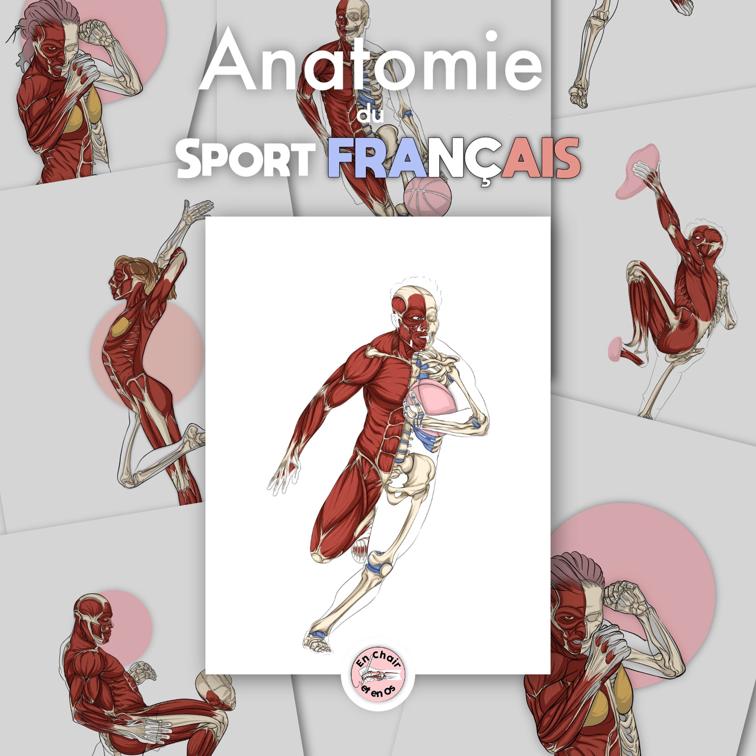
The vertebral column is a central element of the skeleton. Composed of stacked vertebrae, it plays a crucial role in supporting the weight of the human body and protecting the spinal cord, an essential component of our nervous system. The Anatomical Chart: Human Skeleton offers a detailed visual representation of this complex edifice, which is essential for in-depth study. As practitioners, you can use these visual tools to better explain the details of human anatomy to your patients, thereby facilitating their understanding.

Types of bones and classification
Bones can be classified according to their shape and purpose: long, short, flat, and irregular.
Long bones, like the femur or humerus, are essential for mobility, providing the leverage necessary for movement. Conversely, flat bones, like the ribs and skull bones, primarily serve to protect internal organs.
Understanding this classification is crucial for you, as healthcare professionals. For example, a fracture of a flat bone requires a different treatment than that of a short bone. Educational tools such as skeleton models are essential to help you explain these concepts to your patients. This enriches your communication and their understanding.
The functional role of the skeleton
Support and protection
The human skeleton is fundamental for supporting the body. It allows us to stand, walk, run, and move through space. The rib cage, composed of ribs and the sternum, protects vital organs such as the heart and lungs, while the skull protects the brain, one of the most precious organs in our body.
As healthcare professionals, it is crucial to make your patients aware of the importance of this protection. Often, the protection offered by the skeleton is taken for granted until an injury occurs. Using anatomical models during consultations can really help visualize the importance of each element of the skeleton.
Mobility and movement
Another vital function of the skeleton is to enable movement. Thanks to joints, where two bones meet, it is possible to perform a variety of movements. The shoulder and hip joints are key examples of the mobility enabled by the skeleton.
The relationship between the skeleton and muscles is also fascinating. Muscles attach to bones via tendons, thus allowing for a diverse range of movements. Skeleton and musculature models, such as those available on our site En-Chair-et-en-Os, allow for a better understanding of this dynamic. By explaining this to your patients, you show them how muscle strength and bone structure work together to allow them to move.

The importance of the skeleton in the medical field
Diagnosis and treatment of bone pathologies
In your practice, understanding the architecture and role of the skeleton is essential for diagnosing and treating bone-related pathologies. Conditions such as osteoporosis, fractures, or deformities require in-depth knowledge of human anatomy. A fracture of a long bone like the femur can have considerable repercussions on the mobility and quality of life of your patients.
Skeleton models and anatomical posters are indispensable tools for explaining these concepts during consultations. They help you illustrate how injuries affect them and justify recommended treatments. Having a solid understanding of the anatomy and function of each bone allows you to better care for and educate your patients.
Education and awareness
It is also crucial to educate your patients on the importance of bone health. Educational resources like posters and anatomical models play a key role in this process. They allow you to inform your patients about the importance of maintaining healthy bones, encouraging them to adopt preventive behaviors.
Awareness campaigns focused on themes like osteoporosis can greatly benefit from visual aids. These tools make information more tangible and accessible. By using these models, you provide a concrete dimension to your explanations, thereby strengthening your patients' engagement with their health.
Conclusion
The human skeleton is much more than a simple bony framework; it constitutes the very essence of the body. Knowing its composition and role is vital for you, as healthcare professionals. By integrating tools like anatomical posters and skeleton models into your practice, you can enrich your knowledge and improve communication with your patients. Ultimately, a better understanding of skeletal anatomy leads to better health for everyone.
